Seeing Spots or Shadows in Your Vision? Understanding E...
Seeing Spots or Shadows in Your Vision? Understanding Eye Floaters and How They Develop
...
August 18, 2026

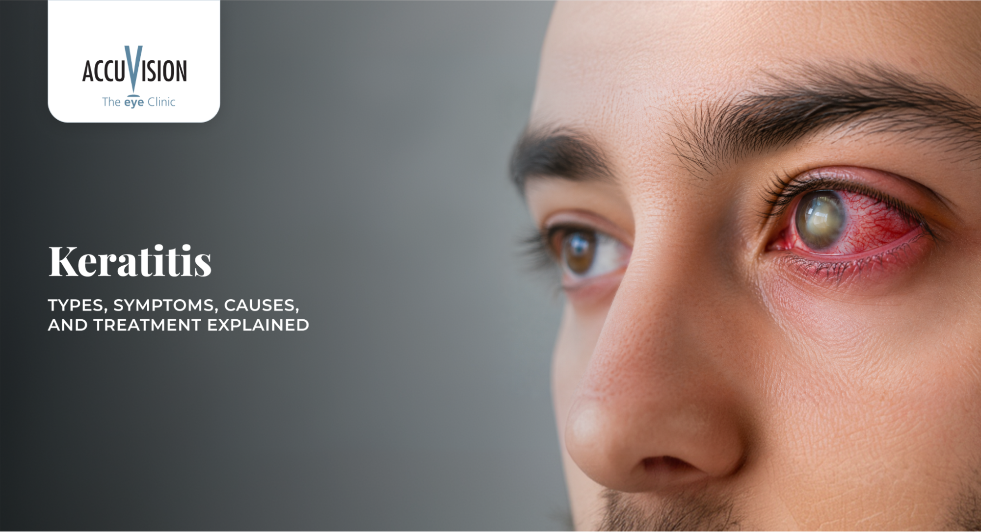
A red, painful eye or sudden blurring of vision can quickly turn from a minor worry into real concern. Many people initially assume these symptoms are due to simple irritation, digital eye strain, or dryness. However, in some cases, they may be early signs of keratitis, an inflammation of the cornea, the clear front window of the eye that plays a vital role in vision.
When keratitis, or a related eye infection or disease, is not assessed and correctly recognised on time, it may lead to complications that affect vision. Understanding keratitis and associated eye conditions, their symptoms, causes, and treatment options can help patients recognise when to seek timely professional care.
In this blog, you will learn about what keratitis is, different types of keratitis, common keratitis symptoms, how microbial keratitis is diagnosed and how keratitis treatment is generally approached by eye care professionals.
Please note that this content serves educational purposes only and does not replace professional medical advice. For sudden, severe or persistent eye symptoms, we recommend booking an urgent eye examination. You can schedule this with the team at AccuVision.
Experiencing Redness or Eye Pain?
Persistent discomfort, redness or sudden blurred vision should be assessed promptly. A comprehensive eye examination at AccuVision can help identify the cause and guide appropriate care.
Keratitis is inflammation of the cornea, the transparent front surface of the eye that helps focus light onto the retina. When the cornea becomes inflamed or infected, it can affect vision and cause discomfort or pain.
Some forms of keratitis may be mild and cause irritation, while others can progress more quickly and may require urgent professional intervention to protect vision.
The organisms responsible for infective keratitis vary considerably by region, largely reflecting differences in climate and environmental exposure.
| Location | Cases (N) | Gram+ Bacteria | Gram− Bacteria | Fungi | Acanthamoeba |
|---|---|---|---|---|---|
| South-west England | 2,614 | 64% | 28% | 7% | 1% |
| Manchester | 4,229 | 63% | 27% | 7% | 2% |
| Ladan, Nepal | 1,897 | 10% (mixed, 39%) | — | 38% | 4% |
In the UK, bacterial infection dominates, with fungal and Acanthamoeba keratitis remaining rare. In Ladan, Nepal, fungal keratitis accounts for over a third of cases, reflecting a broader global pattern where fungal keratitis is more common in tropical, agricultural regions, while bacterial keratitis predominates in higher-income settings, often linked to contact lens wear.
Bacterial keratitis is an infectious condition in which bacteria invade the cornea, often progressing quickly if there is no timely treatment.
Common Risk Factors
Causes
Symptoms to Watch Out For
Fungal keratitis is a corneal infection that may be overlooked or mistaken for other types of infection in the early stages.
Common Risk Factors
Causes
Symptoms to Watch Out For
Acanthamoeba keratitis is a rare but serious corneal infection most commonly linked to contact lens wear and water exposure.
Common Risk Factors
Causes
Symptoms to Watch Out For
Microbial keratitis is a general term for corneal infections caused by different types of microorganisms.
Common Risk Factors
Causes
Symptoms to Watch Out For
Marginal keratitis is an inflammatory condition affecting the outer edge of the cornea and is usually “sterile” rather than a direct or active infection. It may be referred to as a “sterile ulcer”.
Common Risk Factors
Causes
Symptoms to Watch Out For
Superficial punctate keratitis involves tiny surface defects of the cornea caused by dryness or irritation.
Common Risk Factors
Causes
Symptoms to Watch Out For
Viral keratitis is corneal infection or inflammation caused by a virus, most commonly adenovirus, herpes simplex, herpes zoster, or cytomegalovirus.
Common Risk Factors
Causes
Symptoms to Watch Out For
Keratitis is diagnosed by an eye care professional through a comprehensive eye examination, which may include the following tests:
Accurate diagnosis helps guide appropriate keratitis treatment and can reduce the risk of complications such as corneal scarring or vision loss.
| Type of Keratitis | General Treatment Approach* | Key Considerations |
|---|---|---|
| Bacterial keratitis | Typically managed with antibiotic eye drops prescribed by an eye care professional. | In more severe cases, drops may be required more frequently during the initial phase, under professional supervision. |
| Fungal keratitis | Usually managed with antifungal eye drops or medication. | Treatment often requires longer courses and close follow-up by an eye specialist. |
| Acanthamoeba keratitis | Involves specialised anti-amoebic treatments. | May require prolonged therapy and regular monitoring by an ophthalmologist. |
| Superficial punctate keratitis | Commonly managed with lubricating eye drops or gels. | Management may also include addressing dry eye disease, screen habits, or environmental factors as advised. |
| Marginal keratitis | May be managed with anti-inflammatory treatment and eyelid care. | Treating associated eyelid inflammation or blepharitis is often important. |
| Corneal ulcer | Requires urgent specialist assessment and targeted treatment. | Early professional care is essential to reduce the risk of corneal scarring and vision loss. |
*Treatment approaches vary based on individual assessment and should always be guided by a qualified eye care professional.
Consult an eye care professional promptly if you experience:
Contact lens wearers should be particularly cautious, as some forms of microbial keratitis can progress and may cause lasting damage if not treated appropriately.
If you suspect a serious eye problem, or if symptoms are severe or sudden in onset, seek urgent eye care or visit an emergency service as advised in your region.
Keratitis is an inflammatory condition of the cornea that can range from mild irritation to serious infection, making early recognition and professional evaluation essential. Understanding the different types of keratitis, common symptoms, possible causes, and general treatment approaches helps patients seek timely care and reduce the risk of complications. With prompt diagnosis and appropriate management guided by an eye care professional, most cases of keratitis can be treated effectively while protecting long-term vision.
Early evaluation helps protect long-term vision. Schedule a detailed eye examination at AccuVision for accurate diagnosis and personalised guidance.
1. Is keratitis the same as a corneal ulcer?
Not always. Keratitis refers to inflammation of the cornea, while a corneal ulcer is an open sore that can develop in more severe cases of keratitis, such as advanced bacterial or fungal infections. Marginal keratitis can sometimes be called a sterile ulcer. All types of red eye require professional evaluation.
2. Can keratitis go away on its own?
Mild, non-infectious keratitis related to dryness or minor irritation may improve with lubrication. However, infectious keratitis, including bacterial, fungal, or Acanthamoeba keratitis, usually does not resolve on its own and requires professional treatment.
3. Can I shower in my contact lenses?
It’s best avoided. Water contains microorganisms, including Acanthamoeba, that can adhere to lenses and lead to serious infection.
4. Can I swim with my contact lenses?
No, this isn’t recommended, as it significantly increases the risk of Acanthamoeba keratitis and other infections. If swimming is unavoidable, wearing tightly sealed goggles and discarding or thoroughly disinfecting lenses afterwards is advised.
5. Can I sleep in my contact lenses?
Sleeping in lenses reduces oxygen supply to the cornea and weakens its natural defences, raising the risk of bacterial keratitis. Only sleep in lenses specifically approved for overnight wear, and only as directed by your eye care professional.
Stapleton, F. (2023) ‘The epidemiology of infectious keratitis’, The Ocular Surface, 28, pp. 351–363.